Hi, Reader

Welcome to my blog that talk all about radiography and sonography
Showing posts with label Ultrasound on Pregnancy. Show all posts

Sunday, July 22, 2012

Huge and Hyperechoic Yolk Sac on 2D and 3D Ultrasound

Transvaginal ultrasound in the first trimester of pregnancy. Recent improvements in trans-vaginal ultrasound permit the extremely detailed observation of the morphology of the early conceptus in utero. Huge yolk sac on the left : 2D image with normal appearance of early embrio and large yolc sac(arrowheads). On Middle of the figure can see regular heartbeat of 174 bpm. On the Right pic is 3D image of fetus and yolk sac(arrowheads). 8th weeks and 3 days of menstrual age. Fetal demise was confirmed on the next day, Villous chromosome was 48 , xy , + 15, +32, double trisomy.

Hyperechoic yolk sac can you see on the Left with 2D image, On the Right with 3D image. Nine weeks and 1 days of menstrual age. Normal heart beat was seen. Embryonic death was confirmed three days later. Villous chromosome was trisomy 15. Several advantages to ultrasound examination in early pregnancy. Ultrasound performed during the first trimester confirms an intrauterine pregnancy, establishes accurate dating, and is crucial in diagnosing early pregnancy failure and ectopic pregnancy.

Tuesday, June 05, 2012

B-flowmode on Doppler Ultrasound


The B-flowmode is not a Doppler technique based on the processing of the Doppler shift but a B-mode scanning technique that compares gray-scale scans over time to identify changes in the spatial positions of reflectors (blood cells) by the successive emission of coded pulse packets. The echo signals are subtracted from one another, and the brightness is determined by the number of reflectors and partly also by their velocity. The conventional B-scan generated from the stationary echoes is display around the flow information. Since the successive pulses are emitted at defined intervals and in digitally encoded form, it is possible to eliminate interfering echoes and use only the encoded echoes in the subtraction procedure. Hence, only the amplitude signal reflected by moving particles is processed in the interval between two pulses. As the signal strength increases not only with the number of reflecting particles (volume flow) but also with flow velocity, a jet within a stenosis is depicted with a higher signal intensity. The advantage of this technique lies in the simultaneous display in a single image of hemodynamic parameters (comparable to angiography) and morphologic details of the vessel wall with a high resolution while showing no or only little angle dependence. Disadvantages of B-flow imaging are the occurrence of artifacts in highly pulsatile, atherosclerotic vessels, the susceptibility to artifacts caused by wall motion, and the still limited scanning depth. With further technical advancement, B-flow imaging will, in principle, enable morphologic quantification of stenoses and differentiation of the vessel wall from the patent lumen even if only slow flow is present (e.g. in ulceration). The B-mode provides a high-resolution display of the vessel wall contour with separate representation of blood flow in the B-flow mode. There is no superimposition of blood flow information as in color duplex scanning.

Thursday, April 19, 2012

Placenta Development on the Ultrasound Imaging

Primary chorionic will develop between13th and 15th day after the ovulation (during 4th week of gestation) and mark the beginning of the placental development. At the same time, the formation of blood vessels starts in the extraembryonic mesoderm of the yolk sac, the connecting stalk and the chorion. By 18 to 21 days (during the 5th week of gestation), the villi have become branched and the mesenchymal cells within the villi have differentiated into blood capillaries and formed an arteriocapillary venous network. Chorionic villi cover the intire surface of gestational sac until the end of the 8th week. At the time, the villi on the side of the chorion proliferate towards the decidua basalis to form the chorion frondosum, which develops into the definitive placenta. The villi in contact with the decidua capsularis begin to degenerate and form an vascular shell, known as the chorion leave or smooth chorion. The placenta is mostly derived from fetal tissues, when maternal component contributes little to the architecture of definitive placenta.

Normal placentation requires a progressive transformation of the spiral arteries and an infiltration of trophoblastic cells into the placental bed. These physiological changes normally extend into the inner third of the myometrium, and in normal pregnancies, all the spiral arteries are transformed into uteroplacental arteries before 20 weeks of gestation. In some cases of early pregnancy failure and pregnancy-induced hypertention, there is an adequate placentation with a defective transformation of spiral arteries.

3D Power Doppler Studies in Assessment of Early Chorionic Circulation

New developments of the cutting edge of the ultrasound technology enabled us to expand investigations of early placental vascular supply. 3D power Doppler is able to depict the integral 3D image of placenta and embryo and their vascular network. Additionally, it is possible to quantify and express numerically data related to vascular signals in the investigated volume.

Monday, February 13, 2012

The 12th Week of Human Embyo Look by 3D Ultrasound

Characteristic embryological findings: Fetal sex is clearly distinguishableby 12 weeks. The neck is well defined, the face is broad and the eyes are widely separated. By the end 12th weeks, erythropoiesis decreases in the liver and begin in the spleen. The decidua capsularis adheres to the decidua parietalis.
3D ultrasounds findings: Visualization by 3D ultrasounds in the 12th week of gestation enabled more detailed analysis of fetal anatomy, especially limbs. It is possible to count finger and toes. Growing cerebellum is clearly visible. Lateral ventricles dominate the brain.
3D power findings at 11th and 12th week of gestation: with the use of the 3D power dopller, it is possible to depict major branches aorta: common iliac and renal arteries. Circle of willis and it branches are easy visible. From this week, a more detailed anatomical survey can be obtained, including the cerebral and cardiovascular system and the digestive and urinary tracts. At the stage, pulsation of the midlle cerebral artery can be easily discerned as a several vessel. However, until the end of the tenth gestational week, the ultrasonic detected vascular network should be called intracranial circulation. Until the end of the first trimester, the absence of end-diastolic blood flow in fetal and placental components of the circulations is a normal physiological finding.

Sunday, February 12, 2012

3D Ultrasound : 11th Weeks of Pregnancy

The midgut herniation disappear and fetal kidneys produce urine that is excreted into the amniotic fluid.3D ultrasound finding: During the 11th weeks of pregnancy, development of the head and neck continous. Facial details such as nose, orbits, maxilla and mandibles are often visible. Herniated midgut returns into the abdominal cavity. Its persistence after 11th week of gestation is presumptive of an omphalocele. Planar mode enables detailed analysis of the embryonic body with visualization of the stomach and urinary baldder. Kidneys are often visible. Arms and legs continue with development. Hata and coworkers conducted a study an visualization of fetal limbs by 2D and 3D sonography. The ability to visualize fetal hands/finger and feet/toes was better with 3D than with 2D ultrasonography in the late first trimester (detection rate were 65 percent and 41 percent by 3D ultrasonography for hand and feet, respectively and 41 percent and 12 percent, by 2D ultrasonography). Long bones can be visualization as hyperechoic elongated structure inside upper and lower extremities. Detailed 3D analysis of the fetal spine, chest and limbs is obtainable by using the transparent, X-ray like mode. With the use of the transparency X-ray system, starting at 13 week the medular channel, each vertebra and ribs can be visualized and even the intervertebral disks can be measured. This vopens unexpected possibilities for early diagnosis of skeletal malformations.

Friday, February 10, 2012

3D Ultrasound : 9th and 10th Week Embryo Development

Characteristic embryological finding: the head is more rounded and constitutes almost half of the embryo. The hand and feet approach each other. The upper limbs develop faster than the lower limbs, and toward the end of the 9th weeks, the fingers are almost entirely formed. The intestines are in the umbilical cord (Physiological midgut herniation).

3D ultrasound finding: Merz and coworkers were able to provide sticking images of the fetal face at this gestational age. They reported cases in which transvaginal 3D ultrasound produced remarkably well-defined facial image as early as 9 weeks gestation. Sometimes even the external ear can be depicted using 3D surface imaging. Hernation of the midgut is still present as it is a consequence of the rapid growth of the bowel and liver before closure of the abdominal wall. Although this is a physiologic phenomenon, it does not appear in each fetus. Possibly, we cannot, visualize it, or else its size may vary. At 10 weeks, the bowel undergoes to turn of 180 degrees, returning to its original position, at the same time that closure and development of the abdominal wall end. Cerebral hemispheres continue to develop during the 9th and 10th weeks of pregnancy. Visible are lateral ventricles containing hyperechoice choroid plexuses. The head is clearly divided from the body by the neck. External ear is sometimes depicted in the 3D surface image. Herniation of the midgut is present. Dorsal column, the early spine, can be examined in its whole length. The arms with elbow and legs with knees are clearly visible. Feet can be seen approaching the midline.

The size of the lateral ventricles increases rapidly. While the third ventricles is still relatively wide at the beginning of this week, its anteromedial part narrows due to the growth of the thalami. In the fetuses of 25 mm CRL and more, there is a clear gap between the rhombenchepalic and the mesencechepalic is narrow and in most cases, it is not visible in its complete length. The cavity of the dienchepalon decreases in the larger fetuses (CRL = 25mm), and become narrow, especially at its appear anterior part. The spine still characterized by two echogenic parallel lines.

3D power Doppler finding: Fetal structures are now clearly discernible and they are represented by distinct parts of the fetal body-head trunk and limbs. The head measures two-third of the entire body and becomesa distinct anatomical structure. The common and internal carotid arteries may be visualized at the end of the eighth gestational week and the beginning of the ninth week. A cerebral circulation (circle of willis and its major branches) can be documented at eight weeks in the form of discrete pulsation can be detected on tranverse section, lateral on the mesencephalon and chepalic flexsure.

Wednesday, February 08, 2012

3D Ultrasound on Beginning of the 8th Week of Pregnancy


By the beginning of the 8th week, the embryo has develop a skeleton, which is mostly cartilaginous and gives form to its body. The communication between the primitive gut and the yolk sac has been reduced to a relativity small duct (the yolk stalk).

3D ultrasound findings: The most characteristic finding is a complete visualization of the limbs, which end in thicker areas that correspond to the future hands and feet. The shape of the face begins to appear but is not clearly seen. The great majority of embryos show a cranial pole flexion that makes it almost impossible to see the face. Insertion of the umbilical cord is visible on the anterior abdominal wall. During the 8th week of pregnancy, there is expansion of the ventricular system of the brain (lateral, third and midbrain ventricles). Due to these processes, the head erects from the anterior flexion. The vertex is now located over the position of the midbrain. Structures of the viscerocranium are not visible due to their small size. Arms and feet are clearly visible. Insertion of the umbilical cord is visible on the anterior abdominal wall. During the 8th and 9th weeks, the developing intestine is being herniated into the proximal umbilical cord.Blats and coworkers reported on a 7 weeks and 5 days gestational age embryo whose brain structures were analyzed in detail by 3D ultrasound. They described distinct hemispheres how the rhombencephalic cavity (future fourth ventricle) deepens gradually with the growth of the embryos, at the same time decreasing its length. At this time, it has a pyramid-like shape with the central deepening of the pontine flexure as the peak of the pyramid.

3D power Doppler findings: During the 8th and 9th week, developing intestine is being herniated into the proximal umbilical cord, which can be assessed using the technique. By the eighth week, an embryo's length is between 10 and 16 mm, and the CRL is easily measured.

Monday, February 06, 2012

Human Embryo of 7th week


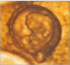
The head is now much larger in relation to the trunk and is more bent over the cardiac prominence. The trunck and neck have begun to straighten. Hand and foot plates are formed and digital or finger rays started to appear.
3D ultrasound findings: During the 7th gestational week, spine gradually becomes visible, as well as limb buds, lateral to the body. Amnion can be seen as a spherical hyperechoic membrane, still close to the embryo. Chorion frondosum can be distinguished from the chorion leave. Fast development of rhombencephalon (hind-brain) occurs. This process gives even more prominence to the head. Head becomes the dominant embryonic structure. Using the multiplanar mode, developing vesicles of the brain can be depicted as anechoic structures inside the head. The biggest, and usually the only visible, is rhombencephalon placed on the top of the head (vertex). The head is strongly flexed anterior being in contact with the chest. The hypoechogenic brain cavities could be identified, including the separated cerebral hemispheres. The lateral ventricles are shaped like small round vesicles. The cavity of the diencephalon (future third ventricle) runs posterior. In the smallest embryos, the medical telencephalon forms a continuous cavity between the lateral ventricles. The future foramen of Monro are wide. In the sagittal plane, the height of the cavity of the diencephalons (future third ventricle) is slightly greater than that of the mesencephalon. Thus, the wide border between the cavities of the diencephalon and the mesencephalon is indicated. The curved tube-like mesencephalic cavity (future sylvian aqueduct) lies anterior, its rostral part pointing caudal. It straightens considerably during the following weeks.
Besides the aorta and umbilical blood flow, at the end of 7th week, 3D power Doppler depicts features of early vascular anatomy on the base of the skull. Vessels are evolving laterally to the mesencephalon and cephalic flexure. Apart from embryonic ciculation, 3D power Doppler can obtain blood flow signals from the intervillous space. The gestational sac occupies about one-third of the uterine volume. The main landmark now is an echogenic fetal pole consisting of embryo adjacent to a cystic yolk sac. The intracranial circulation becomes visble during the seventh week of gestation. At this time, discrete pulsations of the internal carotid arteries are detectable at the base of the skull.

Thursday, January 19, 2012

Basic and Main Component of Colour Doppler Devices

Colour Doppler ultrasound is definitely an ultrasound technique that mixes anatomic information network along with B-mode gray scale as well as color coded blood circulation in two dimensions of period (real time). Here colour is displayed colored, saturation, and lighting to show there is no, path, speed, and type associated with blood flow. Mastery from the basic principles of Doppler, hemodynamic, as well as instrumentation is absolutely necessary to have the ability to produce anatomic and physiologic details are strong.
Colour Doppler ultrasound examination principles
Doppler effect , may be the change in wavelength or even KBS America due to the motion of sound resources, receivers or seem reflector.
In medical ultrasound examination, ultrasound is the supply and receiver transducer that's stationary, while the bloodstream cells, especially red-colored blood cells within the blood stream is a shifting reflector.
Changes in pulse rate of recurrence between the transmitted as well as received ultrasound (Doppler Change) depending on the speed associated with blood flow, flow path relative to the ultrasound examination beam, and the pace of sound surf.

Colour Doppler instrument
You will find three types of devices coluor doppler: Continuous-wave Doppler (CW), Pulse-Doppler duplex (PD / PW), and Color Doppler.
Instruments and PD CW Or PW Doppler blood flow information from the sample volume of the actual CW instrument, sample quantity is relatively large, composed of overlapping regions ultrasound examination beam is released and received through the transducer. In the PD instrument, the dimensions and position could be adjusted and the test volume is sufficiently small less than 1-2 mm. These two instruments present the data in the form of votes and also the Doppler spectrum on the keep track of screen. In the device color doppler, color doppler info obtained from multiple places in a box or even window color as well as presented in two measurements with respect to time.

Colour transition from red-colored to blue or even vice versa showed corrected flow (refereed flow) or even aliasing. Aliasing is an artifact because of doppler shift sample is simply too high or simple rate of recurrence / pulse repeating frequency (PRF) is too low. Once the doppler spectrum, aliasing causes the wrong direction, then colored Doppler color presentation in the opposite direction. Because of aliasing takes place when the Doppler frequency shift surpasses half the PRF is actually color doppler aliasing can be remedied or eliminated through increasing the PRF or transferring the position of the base collection. Here we can see, the utmost speed at both sides of the color club which is the maximum Doppler rate of recurrence shift is equal to fifty percent the PRF, is called the actual Nyquist limit.

To distinguish change flow with aliasing, to think about carefully the restrict on the color alter. Inverted crosses the actual baseline blood flow with the wall so that the filtration system has a transfer restrict of the black, as the intermediate limit aliasing includes a bright, white or even colored in accordance with no more the color bar.

Manage and Optimization associated with Colour Doppler
Control as well as optimization tool colour doppler is very important and must end up being fully mastered prior to performing the evaluation. The main controls for that optimization of color Doppler is: The frequency from the transducer, color bar, colour box, angle, Doppler, colour gain, power result, PRF, Baseline shift, walls filter, ensemble duration, grayscale / colour priority, and persistence.

Rate of recurrence transducer
The selection of transducer frequency is an extremely important decision within the color doppler examination. In the Doppler equation above, it's seen that the Doppler change is proportional to the rate of recurrence emitted, so the greater the transducer frequency the larger the value of doppler shift.

Color Bar
The selection of colour bar / chart depends on the enjoyment of the operator and also the information you want shown. Color map along with variance is mainly employed for detecting the movement echocardiography turbulence.

Colour Doppler and Position boxes
Size, placement and color container angle can be transformed as necessary. Big size and placement can cause the body rate is slower. This really is due to the large size color box leads to the number of scan from the line increases, along with a deeper position may limit the PRF since the time required to transfer and receive replicate doppler longer. Doppler angle place at less than 60 levels.

Colour Gain
Color affects the boosting gain of colour Doppler signal reception. This particular gain must be modified so that the blood flow of knowledge seen by the motion of blood tissue rather than stationary cells.

PRF
PRF primarily affect the level of sensitivity of Doppler and aliasing. Upon color doppler instrument, alterations in PRF is generally done by altering the scale of doppler or even velocity range. This particular effect is always noticed when examining the sluggish venous flow or diastolic arterial movement.

Base Line
Standard shift is used to prevent aliasing without changing the actual PRF or Doppler sensitivity.