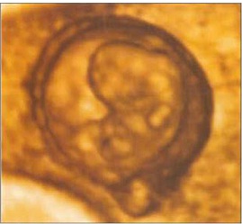
 The head is now much larger in relation to the trunk and is more bent over the cardiac prominence. The trunck and neck have begun to straighten. Hand and foot plates are formed and digital or finger rays started to appear.
3D ultrasound findings: During the 7th gestational week, spine gradually becomes visible, as well as limb buds, lateral to the body. Amnion can be seen as a spherical hyperechoic membrane, still close to the embryo. Chorion frondosum can be distinguished from the chorion leave. Fast development of rhombencephalon (hind-brain) occurs. This process gives even more prominence to the head. Head becomes the dominant embryonic structure. Using the multiplanar mode, developing vesicles of the brain can be depicted as anechoic structures inside the head. The biggest, and usually the only visible, is rhombencephalon placed on the top of the head (vertex). The head is strongly flexed anterior being in contact with the chest. The hypoechogenic brain cavities could be identified, including the separated cerebral hemispheres. The lateral ventricles are shaped like small round vesicles. The cavity of the diencephalon (future third ventricle) runs posterior. In the smallest embryos, the medical telencephalon forms a continuous cavity between the lateral ventricles. The future foramen of Monro are wide. In the sagittal plane, the height of the cavity of the diencephalons (future third ventricle) is slightly greater than that of the mesencephalon. Thus, the wide border between the cavities of the diencephalon and the mesencephalon is indicated. The curved tube-like mesencephalic cavity (future sylvian aqueduct) lies anterior, its rostral part pointing caudal. It straightens considerably during the following weeks.
Besides the aorta and umbilical blood flow, at the end of 7th week, 3D power Doppler depicts features of early vascular anatomy on the base of the skull. Vessels are evolving laterally to the mesencephalon and cephalic flexure. Apart from embryonic ciculation, 3D power Doppler can obtain blood flow signals from the intervillous space. The gestational sac occupies about one-third of the uterine volume. The main landmark now is an echogenic fetal pole consisting of embryo adjacent to a cystic yolk sac. The intracranial circulation becomes visble during the seventh week of gestation. At this time, discrete pulsations of the internal carotid arteries are detectable at the base of the skull.
No comments:
Post a Comment